Blood Vessels Labeled Simple / Q1 Given Alongside Is A Diagram Of Human Heart Showing Its I Lido : In the label the blood vessels activity, students analyze the drawing of the human body and label the arteries that are indicated in the picture.
Blood Vessels Labeled Simple / Q1 Given Alongside Is A Diagram Of Human Heart Showing Its I Lido : In the label the blood vessels activity, students analyze the drawing of the human body and label the arteries that are indicated in the picture.. Terms in this set (6). The lumen is the hollow opening or the space inside the blood vessel. Blood vessels are intricate networks of hollow tubes that transport blood throughout the entire body. Learn vocabulary, terms and more with flashcards, games and other study tools. The inner lining is the endothelium and is surrounded by subendothelial connective tissue.
In the label the blood vessels activity, students analyze the drawing of the human body and label the arteries that are indicated in the picture. Does not form part of the actual practical class based upon the virtual slides. Blood vessels were labeled using qtracker (red). Blood vessels 2 labeled palmar arch digital artery right femoral a right femoral v great saphenous vein left popliteal a right anterior tibial a. August 17, 2020 so, you want to learn.
Chapter 20 Lecture 1224 Hw Flashcards Quizlet from quizlet.com Chapter 14 lecture 1, pages blood vessels blood goes through arteries to arterioles to capillaries to venules to veins into. When chemoreceptors in blood vessels detect high levels of carbon dioxide in the blood, they stimulate all of the following changes except. The iliac, femoral, popliteal and tibial (calf) veins are the deep veins in the legs. Blood vessels cannot function properly when inhibited by vascular diseases. The inner lining is the endothelium and is surrounded by subendothelial connective tissue. The course of the abdominal aorta is quite simple. The most important types, arteries and veins, carry all blood vessels have the same basic structure. Does not cover the pathology content.
Through the thin walls of the capillaries, oxygen and nutrients pass from blood into tissues, and waste products.
Blood vessels are an integral component of the circulatory system. Arterioles connect with even smaller blood vessels called capillaries. To print or download this file, click the link below Each pixel must be labeled 1 if it is part of a blood vessel in the image, and 0 if not. The intima is a simple epithelium made up of a single layer of flat epithelial cells. Structure of blood vessels in general, blood vessels have a walls composed of three layers as follows: Through the thin walls of the capillaries, oxygen and nutrients pass from blood into tissues, and waste products. Dimitrios mytilinaios md, phd • last reviewed: Blood vessels flow blood throughout the body. Veins (in blue) are the blood vessels that return blood to the heart. August 17, 2020 so, you want to learn. Chapter 14 lecture 1, pages blood vessels blood goes through arteries to arterioles to capillaries to venules to veins into. Retinal_blood_vessel_segmentation.ipynb this is a simple implementation neural net with.
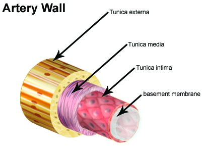
Blood vessels are intricate networks of hollow tubes that transport blood throughout the entire body. Blood vessels are referred to collectively as the vascular system and, together with the heart, make up the circulatory system or cardiovascular system. Which of the labeled layers in the diagram of the arterial wall is composed of a simple squamous epithelium, a basement membrane and a layer of. Terms in this set (6). Vessels that carry blood away from heart, surrounded by thick layer of smooth muscle, high levels of bp review:
Seer Training Classification Structure Of Blood Vessels from training.seer.cancer.gov Does not form part of the actual practical class based upon the virtual slides. Chapter 14 lecture 1, pages blood vessels blood goes through arteries to arterioles to capillaries to venules to veins into. In the label the blood vessels activity, students analyze the drawing of the human body and label the arteries that are indicated in the picture. Blood vessels are referred to collectively as the vascular system and, together with the heart, make up the circulatory system or cardiovascular system. Start studying blood vessels labeling. All blood vessels are specifically structured to perform their function. The most important types, arteries and veins, carry all blood vessels have the same basic structure. To print or download this file, click the link below
Retinal_blood_vessel_segmentation.ipynb this is a simple implementation neural net with.
Chapter 14 lecture 1, pages blood vessels blood goes through arteries to arterioles to capillaries to venules to veins into. The blood vessels are the components of the circulatory system that transport blood throughout the human body. The capillaries also connect the branches of arteries and to. Blood vessels are referred to collectively as the vascular system and, together with the heart, make up the circulatory system or cardiovascular system. Simple squamous epitheliumendothelium in blood vessels blood vessel color images The course of the abdominal aorta is quite simple. This page is about blood vessel histology slide labeled,contains artery microscope slide. The lumen is the hollow opening or the space inside the blood vessel. Blood and lymph vessels arteries and nerves of hand: Each pixel must be labeled 1 if it is part of a blood vessel in the image, and 0 if not. Blood vessels are intricate networks of hollow tubes that transport blood throughout the entire body. Blood vessels labeled simple : Ready to learn about the blood vessels of the abdomen and pelvis (the abdominopelvic blood vessels)?
Blood vessels are intricate networks of hollow tubes that transport blood throughout the entire body. Blood vessels were labeled using qtracker (red). The course of the abdominal aorta is quite simple. It is made up of. Blood travels from the heart in arteries, which branch into smaller and smaller vessels, eventually becoming arterioles.
Interlobular Artery Anatomy Britannica from cdn.britannica.com The blood vessels are the components of the circulatory system that transport blood throughout the human body. Blood vessels are flexible tubes that carry blood, associated oxygen, nutrients, water, and hormones throughout the body. Blood vessels are referred to collectively as the vascular system and, together with the heart, make up the circulatory system or cardiovascular system. The course of the abdominal aorta is quite simple. All vessels feature varying lumen size. (no rating) 0 customer reviews. Terms in this set (6). The best websites voted by users.
This page is about blood vessel histology slide labeled,contains artery microscope slide.
(no rating) 0 customer reviews. These vessels transport blood cells, nutrients, and oxygen to the tissues of the body. August 17, 2020 so, you want to learn. Hma practical 3 virtual slides. Structure of blood vessels in general, blood vessels have a walls composed of three layers as follows: Blood vessels are intricate networks of hollow tubes that transport blood throughout the entire body. The inner lining is the endothelium and is surrounded by subendothelial connective tissue. Blood and lymph vessels arteries and nerves of hand: Arterioles connect with even smaller blood vessels called capillaries. Hma practical 3 for monday july 23 and wednesday july 25. Blood vessels cannot function properly when inhibited by vascular diseases. One of the most common diseases of the arteries is called atherosclerosis. Vessels that carry blood away from heart, surrounded by thick layer of smooth muscle, high levels of bp review:
One of the most common diseases of the arteries is called atherosclerosis blood vessels labeled. Veins (in blue) are the blood vessels that return blood to the heart.


0 Comments